Muscles Of The Torso / Back Muscles Torso Leyton Sports Massage / This image shows the muscles of our body and displays them on both male and female diagram showing:
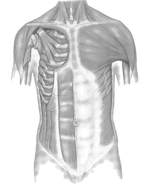
Muscles Of The Torso / Back Muscles Torso Leyton Sports Massage / This image shows the muscles of our body and displays them on both male and female diagram showing:. Extending across the anterior surface of the body from the superior border of the pelvis to the inferior border of the ribcage are the muscles of the abdominal wall, including the transverse and rectus abdominis and the internal and external obliques. They are most prevalent in the supply of skin covering the broad, flat muscles of the torso (e.g., latissimus dorsi, rectus abdominis). Why is my entire torso cramping? The action refers to the action of each muscle from the standard anatomical position. Related posts of muscles of the torso diagram muscle anatomy ribs.
The torso also harbours many of the main groups of muscles in the body, including the: The biceps brachii, deltoid, external oblique, infraspinatus, inguinal ligament, internal oblique, latissimus dorsi, levator scapulae, pectoralis major, platysma, rectus abdominus, rhomboid major & minor,. Every effort was made to make sure this is anatomically correct as possible. If unilateral innervation of the rectus abdominis muscle occurs, it is also capable of lateral flexion of the torso. One of the large muscles of the leg, it connects to the heel.

Extending across the anterior surface of the body from the superior border of the pelvis to the inferior border of the ribcage are the muscles of the abdominal wall, including the transverse and rectus abdominis and the internal and external obliques.
When employed unilaterally the muscle functions to tilt the torso toward that side of the body and/or to rotate the torso away from that side of the body (contralateral rotation. During active innervation by the subcostal Pectoral muscles abdominal muscles lateral muscle epaxial muscles This lecture describes four groups of muscles attached to the skeletal structures of the torso. Bilaterally this muscle extends the torso. They cover areas of the body and get their name from them (fascia of the shoulder, chest, thigh, forearm, etc.). Every effort was made to make sure this is anatomically correct as possible. The rectus abdominis muscle is also known as the abs. The biceps brachii, deltoid, external oblique, infraspinatus, inguinal ligament, internal oblique, latissimus dorsi, levator scapulae, pectoralis major, platysma, rectus abdominus, rhomboid major & minor,. Its fibers are concentrated at the sides of the abdomen and, like the external oblique, has an aponeurosis covering the medial abdomen under the rectus abdominis. Although the torso rotation movement targets your obliques, several other muscles, including quadratus lumborum, rectus abdominis and transverse abdominis, engage to assist your core and stabilize your torso. Key parts of your spine include vertebrae (bones), disks, nerves and the spinal cord. Muscle tissue consists of all skeletal muscles:
Bilaterally this muscle extends the torso. The anterior muscles of the trunk (torso) are associated with the front of the body, include chest and abdominal muscles. The semispinalis dorsi and semispinalis capitis muscles also extend the back. This video is about muscles of the torso. The rectus abdominis muscle is also known as the abs.

Another common cause of muscle spasms on the left side of the torso is irritable bowel syndrome (ibs).
Pyramidal muscle the pyramidal muscle originates from the superior margin of the pubic bone and the symphysis, and is located anterior to the rectus abdominis muscle. For more videos visit seewhayanatomy.com or follow us on twitter @seewhyanatomy The deepest layer has the transverse abdominis muscle, whose fibers run laterally. Although the torso rotation movement targets your obliques, several other muscles, including quadratus lumborum, rectus abdominis and transverse abdominis, engage to assist your core and stabilize your torso. Muscles of the torso medical edition each muscle of the torso is textured and has the correct origin and insertion points. The muscles of the human body can be categorized into a number of groups which include muscles relating to the head and neck, muscles of the torso or trunk, muscles of the upper limbs, and muscles of the lower limbs. The disks that cushion vertebrae may compress with age or injury, leading to a herniated disk. It is strapped shaped and winds across the front of the thigh, from the hip to the inner side of the tibia. Again, this view of the muscles of the torso seen from the side is obviously informed by dissection, even though these are superficial muscles which show clearly under the surface of the skin. Also capable of lateral flexion of the torso. Key parts of your spine include vertebrae (bones), disks, nerves and the spinal cord. In the upper back region, the trapezius, rhomboid major, and levator scapulae muscles anchor the scapula and clavicle to the spines of several vertebrae and the occipital bone of the skull. It is inserted in the linea alba and runs within the rectus sheath.
It flexes and extends the foot, ankle, and knee. Muscles of the chest pectoral muscles Extensor muscles of the hand 6. Muscles fibers originating from two to four vertebrae below connect to the spinous process of each vertebrae except c1. The biceps brachii, deltoid, external oblique, infraspinatus, inguinal ligament, internal oblique, latissimus dorsi, levator scapulae, pectoralis major, platysma, rectus abdominus, rhomboid major & minor,.
Pulls arm anteriorly and across chest, rotates humerus, or add….
During active innervation by the subcostal If unilateral innervation of the rectus abdominis muscle occurs, it is also capable of lateral flexion of the torso. The biceps brachii, deltoid, external oblique, infraspinatus, inguinal ligament, internal oblique, latissimus dorsi, levator scapulae, pectoralis major, platysma, rectus abdominus, rhomboid major & minor,. Muscles fibers originating from two to four vertebrae below connect to the spinous process of each vertebrae except c1. The action refers to the action of each muscle from the standard anatomical position. It flexes and extends the foot, ankle, and knee. In the upper back region, the trapezius, rhomboid major, and levator scapulae muscles anchor the scapula and clavicle to the spines of several vertebrae and the occipital bone of the skull. These muscles work together to move the scapula anteriorly and laterally during pushing, throwing, or punching motions. It flexes and extends the foot, ankle, and knee. This image shows the muscles of our body and displays them on both male and female diagram showing: The dominant muscle in the upper chest is the pectoralis major. This arrangement is most common between the longer, thinner muscles of the extremities (e.g., radial forearm flap, dorsalis pedis flap). The small muscles of the vertebrae (the multifidi and rotators) help rotate, extend, and side bend the back.


Komentar
Posting Komentar